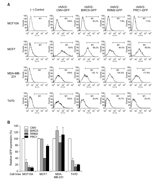
Figure 4.
The GFP expression in rAAV2-infected breast normal and cancer cell lines. The promoter activity of the rAAV2 vector containing the GFP reporter gene under cancer-specific promoters of PRC1, RRM2 or BIRC5 was compared to that under the CMV promoter in breast normal (MCF10A) and cancer (MCF7, T47D and MDA-MB-231) cell lines. The cells were seeded at 1 × 10⁴ cells/well in 12-well plates and infected with rAAV2 vectors of 2 × 10⁵ MOI for 72 h. The GFP expressions in the transduced cells were measured using a FACS flow cytometry and the experiment was repeated three times in triplicate. (A) One of representative FACS data collected from 10,000 cells/sample was presented in a histogram of fluorescence intensity (X-axis) and cell counts (Y-axis). The fluorescence intensity of the (-) control cell of mock infection was used to distinguish the rAAV-infected population from the uninfected population by setting M1 for each tested cell line, since the rAAV-infected cells emitted a fluorescent signal ranging M1. (B) The relative GFP expression. The GFP expression in each case was normalized by transduction efficiency of each rAAV measured by semi-quantitative PCR of AAV genomic DNA. The GFP expression derived from rAAV2-CMV-GFP was considered to be 100, and relative GFP expressions in the infected cells with rAAV2 containing the PRC1, RRM2 or BIRC5 promoter were plotted in the bar graph. The average values of the independently repeated experiments were applied and plotted in the graph.

