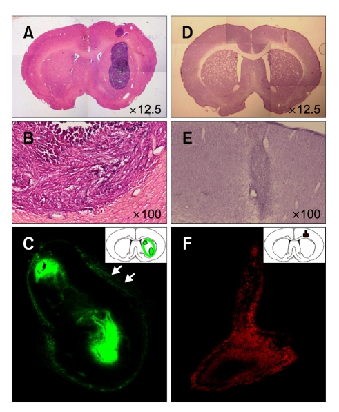
Figure 1.
Infiltrating nature of C6 glioma and NSC HB1.F5 in normal brain. A-C: SD rat was stereotactically injected with Vybrant® DyeCycle™ Green-stained C6 rat glioma cells in the right striatum. Five days later, the right cortex of the animal received another injection with HBSS to emulate minor trauma in CNS. Seven days later, the brain sections of the recipient were observed. C6 rat glioma cells were developed to form a huge mass, occupying almost the entire striatum. (A and B) Hematoxylin and eosin-stained and (C) DyeCycle-labeled C6 glioma. D-F: SD rat received the 1st injection of C6 cell-free HBSS in the right striatum; five days later, the right cortex of the animal received a 2nd stereotaxic injection of Dil-labeled HB1.F5 cells. The brain sections of the recipient were then assessed. Whole NSCs remained at the original injection site as long as a week. (D) Hematoxylin and eosin-stained, and (E and F) Dil-labeled HB1.F5 cells.

