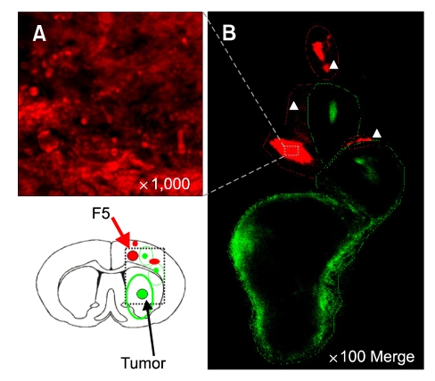
Figure 4.
NSCs approached glioma cells in the vicinity of NSC mass. Dil-labeled HB1.F5 cells were infused into the rodent brain previously implanted with DyeCycle™-labeled C6 glioma cells. Three days later, recipients were sacrificed and their brain sections processed to detect fluorescing cells. Most NSC localized near the transplanted site (red arrows in left lower panel). Extensive migration of Dil-labeled red HB1.F5 was noted in the vicinity of the green-stained infiltrating glioma (as demarcated by the white arrow in B). Green: DyeCycle green-labeled C6, Red: Dil-labeled NSCs (A) High resolution (1,000 ×) of transplanted NSC. (B) 100 × merged image.

