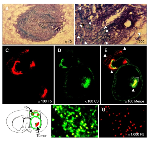
Figure 5.
NSCs progressively invade the tumor core, surrounding the glioma mass. Dil-labeled HB1.F5 cells were infused into the rodent brain previously implanted with DyeCycle™-labeled C6 glioma cells. Five days later, recipients were sacrificed and their brain sections processed for observation. C6 rat glioma cells continued to disseminate themselves through the entire striatum of the recipient's brain and their mass enlarged to reach almost to the brain surface. X-gal-stained blue NSCs were located in virtual juxtaposition along the border of the C6 tumor mass stained by hematoxylin where the tumor interfaced with normal tissue that was being aggressively infiltrated (as indicated by white arrowheads in B). Several NSCs were also found invading deeply into the tumor mass and up to the core and intermixing with tumor cells as shown in the merged images (E and F). In the cortex, NSCs appeared to surround the tumor mass and to prevent the tumor cells from moving into the normal brain tissue. A few NSCs pursued the solitary tumor cells that escaped from the cerebral cortex (B and E). It was particularly noteworthy that numerous NSCs reached the tumor core, far from the original injection site and intermixed with the tumor cells as shown in the merged images and noted by the white arrowhead in B, E and F. (A) Low-resolution (40 ×) and (B) High-resolution images of Hematoxylin-stained C6 and X-gal stained NSC. (C-E) Low-power and (F and G) High-power confocal images. (E and F) merged images. Green: DyeCycle-labeled C6, Red: Dil-labeled NSC.

