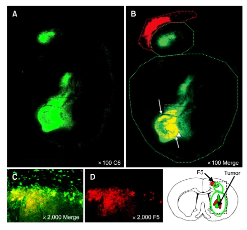
Figure 6.
NSCs were distributed throughout the tumor bed. Dil-labeled HB1.F5 cells were infused into rodent brain previously implanted with DyeCycle™-labeled C6 glioma cells. Seven days later, recipients were sacrificed and their brain sections were processed to detect fluorescing cells. NSCs invaded the tumor bed until they were mixed throughout the tumor bed that occupied the whole striatum and cortex, especially in the tumor core as noted by the white arrow in B. (A and B) Low-power (100 ×) and (C and D) High-power (2,000 ×) confocal images. (B and C) Merged images of DyeCycle-labeled C6 and Dil-labeled NSCs. Green: DyeCycle-labeled C6, Red: Dil-labeled NSC.

