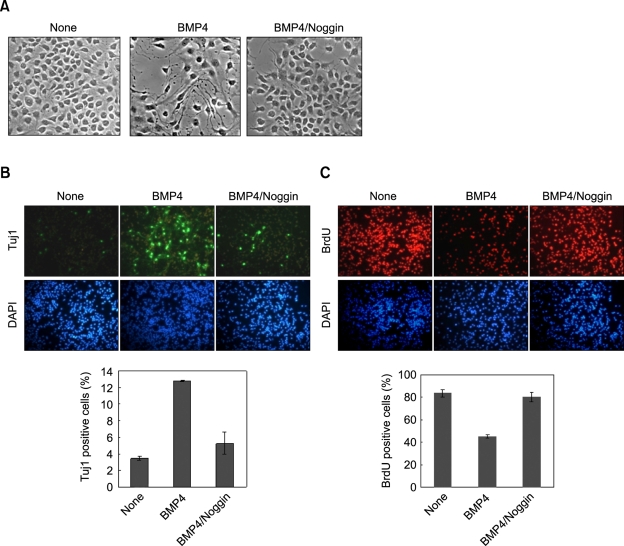
Figure 1.
Effect of BMP4 on differentiation and proliferation of NSCs. NSCs that were grown in the N2 medium containing bFGF (10 ng/ml) were treated with 20 ng/ml of BMP4 for 48 h. One case required noggin was co-treated with BMP4 (150 ng/ml). (A) Micrographs were taken, using a phase-contrast microscope at 400× magnification. (B-C) The cells were processed for immunofluorescent labeling of Tuj1 (green) and BrdU (red). The nuclei were counterstained with DAPI (blue). All of the images taken of the cells labeled with fluorescent chemicals were magnified 200× using a fluorescent microscope. The percentages of BrdU-positive and Tuj1-positive cells were determined. The error bars indicate the standard deviations of three independent experiments. The data represent the mean ± SD of three separate experiments.

