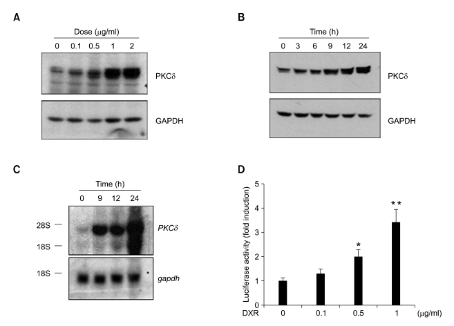
Figure 2.
Up-regulation of PKCδ expression by doxorubicin (DXR). (A, B) L1210 cells were treated with the indicated concentrations of DXR for 12 h (A) or with 1 µg/ml DXR for the indicated lengths of time (B). Total cell lysates were prepared and subjected to Western blotting with anti-PKCδ antibody (upper panels). The same blot was re-probed with anti-gapdh antibody as an internal control (lower panels). The blots shown are representative of results obtained in three independent experiments. (C) Total RNA was isolated from cells treated as in (B) and subjected to Northern blotting. The blot was hybridized with a 32P-labeled PKCδ cDNA probe (upper panel), stripped, and re-probed with a 32P-labeled GAPDH probe (lower panel). The blots shown are representative of results obtained from two independent experiments. (D) Sub-confluent NIH 3T3 cells grown in 12-well plates were transfected with 0.5 µg of mPKCδ-Luc(-1192/+10) along with 5 ng of pRL-null vector. Twenty-four hours after transfection, the cells were treated with DXR (0.1, 0.5, or 1 µg/ml) for 8-12 h. Firefly luciferase activity was normalized to Renilla luciferase activity. The data shown represent the mean ± S.D. (error bars) of three independent experiments performed in triplicate. The statistical significance of the assay was evaluated using Student's t-test (*, P < 0.05; **, P < 0.01 compared with untreated control).

