Figure 3.
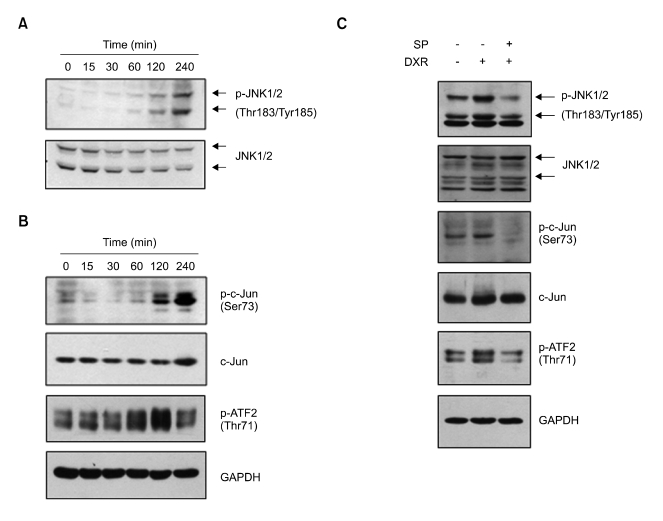
DXR activation of the JNK pathway in L1210 cells. (A) L1210 cells were serum-starved (grown in 0.5% serum) for 24 h and then exposed to 1 µg/ml DXR for the indicated lengths of time up to 240 min. Total cell lysates were prepared and subjected to Western blotting with anti-phospho-JNK1/2 (Thr183/Tyr185) antibody. The blot was stripped and re-probed with anti-total JNK antibody as an internal control. (B) L1210 cells were treated as in (A). Total cell lysates were subjected to Western blotting with anti-phospho-c-Jun (Ser73) and anti-phospho-ATF2 (Thr71) antibodies. The blots were re-probed with antibodies against total c-Jun and GAPDH as internal controls. (C) L1210 cells were treated with 0 or 40 µM SP600125 (SP) for 30 min and then treated with 0 or 1 µg/ml DXR for 2 h as indicated. Total cell lysates were prepared and subjected to Western blotting with antibodies specific for phospho-JNK1/2 (Thr183/Tyr185), -phospho-c-Jun (Ser73), or phospho-ATF2 (Thr71). The blots were stripped and re-probed with anti-JNK, -c-Jun, or -GAPDH antibody. Blots shown are representative of at least three separate experiments.

