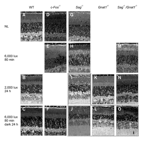
Figure 1.
Retinal sections from mice after exposure to light for various time periods. The dot box indicates severe cell damage. Some pictures taken under certain conditions and at different time points where morphological changes did not occur were not included. NL, no light; WT, wild type; Sag-/-, arrestin knockout; Gnat1-/-, transducin knockout; Sag-/-/Gnat1-/-, arrestin/transducin double knockout; c-Fos-/-, c-Fos knockout.

