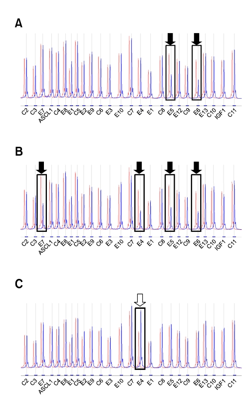
Figure 2.
Multiplex ligation-dependent probe amplification (MLPA) electrophoresis tracings for normal control (red-colored) and PKU subjects (blue-colored) with exon deletions or duplication. The boxes and arrows indicate decreased or increased patient's peaks relative to the control's peaks. A. Decreased MLPA tracing of the PAH gene peaks from a PKU patient with a deletion of exons 5 and 6 (filled arrows). B. Decreased MLPA tracing of the PAH gene peaks from a PKU patient with a deletion extending from exons 4 to 7 (filled arrows). C. Increased MLPA tracing of the PAH gene peaks from a PKU patient with a duplication of exon 4 (open arrow). C, control; E, exon

