Fig. 3.
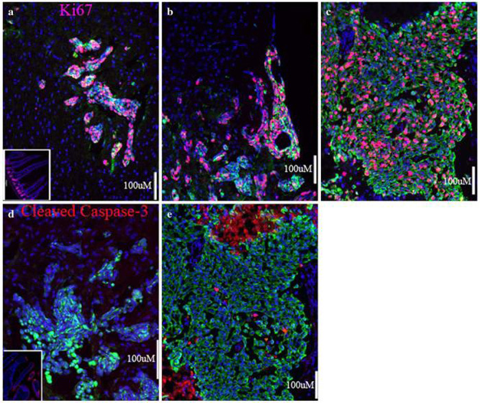
Life and death in breast cancer brain metastases. (a) A cluster of 231-BR cell metastases in the mouse cerebral cortex. Cycling cell nuclei, expressing Ki67, appear pink. Little to no proliferation is visible in the surrounding brain tissue. Inset in (a) shows positive-control staining with anti-Ki67 in the crypts of mouse intestine. (b) A meningeal metastasis at the surface of the brain shows levels of proliferation similar to that of parenchymal metastases. (c) A surgical sample from a brain metastasis of ductal carcinoma showing many Ki67-positive cycling tumor cells. (d) Apoptosis is rare in the xenograft model, as is apparent from the lack of staining for Cleaved Caspase-3 (red). Inset in (d) shows positive-control staining for Cleaved Caspase-3 in the villi of mouse intestine. (e) A few apoptotic cells are visible among the proliferating carcinoma cells from a surgical specimen of brain metastasis of ductal carcinoma. Necrotic areas, at the top and bottom left of (e) also show reactivity to the anti-Cleaved Capsase-3 antibody; (c) and (e) are from neighboring sections of the same metastasis. Five micron (human) or ten micron (mouse) sections were prepared from frozen samples and fixed with paraformaldehyde. Cytokeratin-positive tumor cells are stained green in all panels, and all nuclei counterstained with DAPI in blue. Scale bars indicate 100 micrometers

