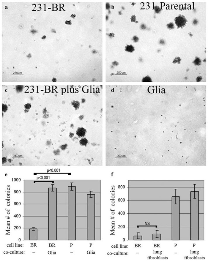
Fig. 6.
Glial cells from mouse cerebral cortex stimulate anchorage-independent growth of 231-BR cells in vitro. (a) Colonization by 231-BR cells alone for 14 days in soft agar. (b) Colonization by 231-Parental (lung metastatic) cells alone for 14 days in soft agar. (c) Increased colonization of 231-BR cells in co-culture with mixed glial cells for 14 days in soft agar. (d) Glial cells alone for 14 days in soft agar. (e) Quantification of a typical experiment, showing the effect of glia on 231-BR growth in soft agar. Mean number of colonies (>50 μm) from triplicate samples ± standard deviation are shown. Results of post-hoc Tukey-Kramer Multiple Comparisons Testing are shown on the graphs. BR = 231-BR (brain metastatic) cells; P = 231-Parental (lung metastatic) cells. (f) MRC-5 lung fibroblasts do not stimulate 231-BR cell growth in soft agar (NS = not significant)

