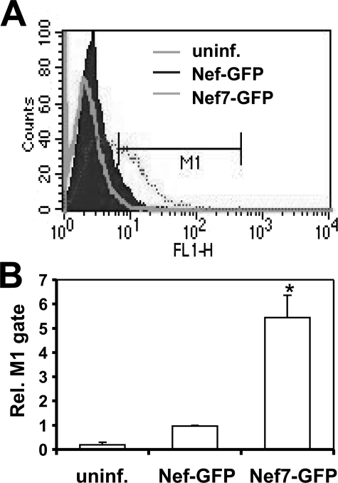
FIGURE 2.
Delivery of virion WT Nef and Nef7 into cells. 293T cells were transfected with HIV.env–.nef– and VSV-G and with WT Nef-GFP or Nef7-GFP expression plasmids. Progeny viruses were collected and quantitated as stated above. Viruses with an equal level of RT activity were used to infect fresh 293T by spinoculation. Uninfected cells (uninf.) were used as a control. After 3 h cells were washed to remove unbound viruses and then trypsinized to remove cell surface-bound virus. These cells were then analyzed for GFP-positive cells by fluorescence-activated cell sorter to detect intracellular delivery of virion Nef (A). FL1-H, increasing GFP intensity. The relative level of GFP-positive cells from three independent experiments was calculated with the number of GFP-positive cells in WT Nef-GFP infection set to 1 (B).

