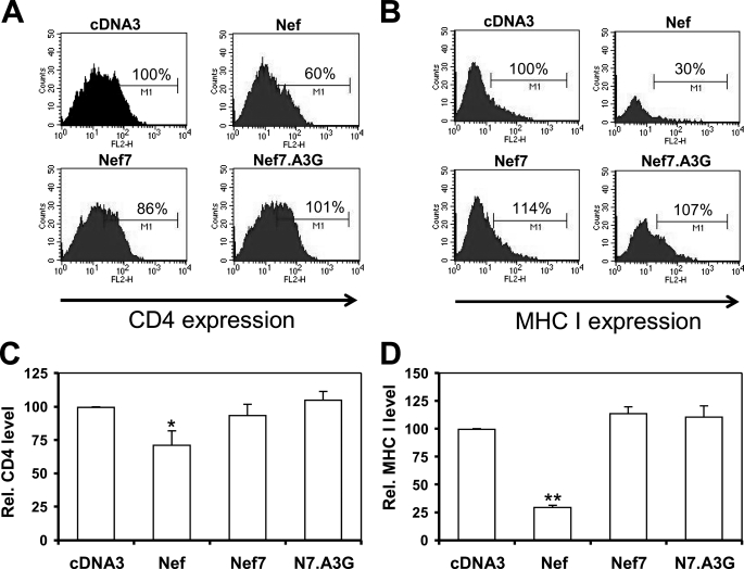
FIGURE 5.
Effects of Nef, Nef7, and Nef7.A3G on cell surface expression of CD4 or MHC I. HeLa cells were transfected with CD4, GFP, and Nef, Nef7, or Nef7.A3G expression plasmids with pcDNA3 included as a control. At 24 h cells were harvested and stained with an anti-CD4 antibody followed by a phycoerythrin-conjugated secondary antibody. The cells were then gated for the GFP+ cells by fluorescence-activated cell sorter, and only the GFP+ cells were analyzed for cell surface CD4 expression (A). WB, Western blot. Similarly, HeLa cells were transfected with HLA-A2, GFP, and Nef, Nef7, or Nef7.A3G expression plasmids and analyzed for cell surface MHC I expression (B). Cell staining without anti-CD4 or anti-MHC I antibodies was included as the negative control to ensure that these secondary antibodies did not have nonspecific staining (data not shown). The relative CD4 and MHC I levels from three independent experiments were calculated with the CD4 or MHC I level in pcDNA3-transfected cells set to 100% (C and D).

