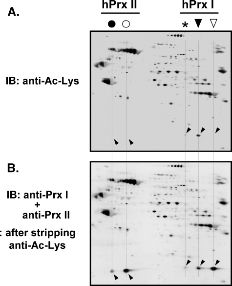
FIGURE 2.
N-Acetylation of the acidic migrated hPrx I in HeLa cells. HeLa cell lysate (50 μg) was separated on a two-dimensional polyacrylamide gel, followed by immunoblotting with anti-acetylated lysine antibody (anti-Ac-Lys; Cell Signaling Technology). For accurate control of relative sensitivity, alkaline phosphatase-conjugated anti-rabbit IgG with nitro blue tetrazolium/5-bromo-4-chloro-3′-indolyl phosphate was used for the visualization of immune complexes (A). After mild stripping of the antibodies, as described in the manufacturer's protocol (Abcam), the membranes were reprobed with a mixture of anti-Prx I and anti-Prx II antibodies (B). The spot positions of acidic migrated (▾) and reduced (▿) forms of hPrx I and acidic migrated (•) and reduced (○) forms of hPrx II are indicated. An asterisk indicates the further acidic migrated positions of hPrx I. Spots that cross-reacted with anti-Prx I and anti-Prx II are indicated with slanted downward and slanted upward arrowheads, respectively. IB, immunoblot.

