FIGURE 5.
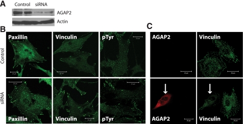
Knockdown of AGAP2 stabilizes focal adhesions. A, expression of endogenous AGAP2 in U87 cells was suppressed using siRNA, and the nontargeting siRNA was used as a control. Knockdown of AGAP2 was confirmed by Western blotting, and actin was examined as the protein loading control. B, visualization of focal adhesions in U87 cells with and without AGAP2 knockdown. Focal adhesions were stained using anti-paxillin, anti-vinculin, or anti-phosphotyrosine (pTyr) antibodies in control (top panels) and AGAP2 knockdown (bottom panel) cells. Scale bar, 20 μm. C, rescue of AGAP2 knockdown. Expression of endogenous AGAP2 in U87 cells was suppressed by shRNA targeting the 3′-untranslated region. Plasmids encoding empty vector (top panels) or FLAG-AGAP2 (bottom panels) were transfected into U87 cells with stable knockdown of AGAP2. Focal adhesions were examined by staining with anti-vinculin antibody. Arrow indicates the cell expressing FLAG-AGAP2.

