FIGURE 5.
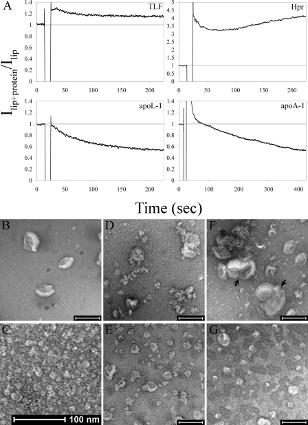
TLF and its protein components exhibit dramatically different membrane altering activities. A, the 90° light-scattering intensity of susceptible liposomes (lip) was monitored as an indicator of relative particle size at pH 5.0 with the addition (indicated by the drop in signal) of 20 nm TLF (10μg/ml), Hpr (0.9μg/ml), apoL-1 (0.84 μg/ml), or 60 nm apoA-1 (1.68 μg/ml). B–G, transmission electron micrographs of negatively stained preparation of soy bean azolectin liposomes (B), TLF alone (C), liposomes treated with TLF (D), liposomes treated with apoL-1 (E), liposomes treated with Hpr (F), and liposomes treated with apoA-1 (G). Scale bars are 200 nm, except in C. The liposomes in D appear decorated with TLF particles. Apparent fusion products and agglutinated liposomes in F are indicated by an arrow and asterisk, respectively.

