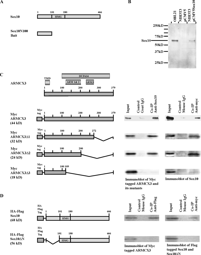
FIGURE 1.
Identification of ARMCX3 as a Sox10-interacting protein. A, schematic representation of the rat Sox10 protein indicating the HMG box (top figure). Numbering refers to amino acids. A cDNA fragment that encodes the first 100 amino acids of Sox10 was subcloned into pGBKT7 to yield the bait plasmid pGBKT7-Sox10N100 (lower figure). B, detection of endogenous Sox10 in OBL21 whole cell extracts and exogenous Sox10 in whole cell extracts of pCMV5-Sox10-transfected NIH3T3 cells. Parallel cultures of pCMV5-transfected NIH3T3 cells were analyzed on the same Western blot. C, schematic representation of ARMCX3 indicating the three ARM repeats, the putative transmembrane domain (TMD) and the DUF634 domain (top figure). Myc-tagged ARMCX3 and its three deletion constructs are shown in the lower portion of the figure. Cell lysates were prepared from OBL21 cells transfected with pCMV-Myc-ARMCX3 and the three deletion constructs. Co-IPs were done using anti-Sox10 and anti-Myc antibodies, as indicated. Mouse IgG and goat IgG were used as negative controls. The left immunoblot presents the results from a precipitation with anti-Sox10 followed by blotting with anti-Myc while the right immunoblot presents the reciprocal experiment in which anti-Myc was used for immunoprecipitation and anti-Sox10 was used for the immunoblotting. D, pCMV-Myc-ARMCX3 was co-transfected with either pCMV-HA-FLAG-Sox10 or pCMV-HA-FLAG-Sox10ΔN into Neuro-2A cells. Cell lysates were prepared, and Co-IPs were done using anti-FLAG and anti-Myc antibodies.

