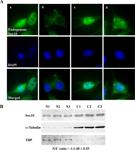
FIGURE 2.
Subcellular localization of Sox10. A, endogenous Sox10 was detected in OBL21 cells by immunofluorescence. Nuclei were stained with 4′,6-diamidino-2-phenylindole (DAPI). The four sets of images show nuclear (a), cytoplasmic (b), and nucleocytoplasmic (c and d) localization of Sox10. B, nuclear and cytoplasmic fractions were isolated from OBL21 cells. Successful isolation of nuclear protein was verified by immunoblot analysis of TBP while successful isolation of cytoplasmic protein was confirmed by immunoblot analysis of α-tubulin. Sox10 was detected by immunoblot analysis using anti-Sox10 antibody. The Sox10 nuclear:cytoplasmic (N/C) ratio was determined by semi-quantitative immunoblot analysis (see “Experimental Procedures”). The results of three independent experiments are shown.

