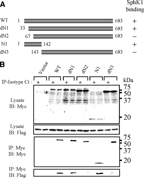
FIGURE 4.
Mapping of the SphK1-binding site on BVDV NS3. A, schematic representation of NS3 and its deletion mutants. B, SphK1 expression vector in combination with expression vectors of Myc-NS3 and its deleted mutants were cotransfected into LB9.K cells. Proteins immunoprecipitated (IP) with anti-Myc mAb (even lanes) or isotype control IgG (Isotype Ct; odd lanes) were subjected to Western blotting (IB) using anti-FLAG mAb. Data shown in each panel are representative of at least three independent experiments.

