FIGURE 4.
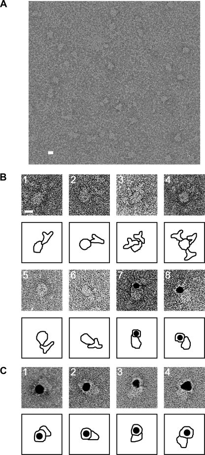
Electron microscopy of negatively stained Orai1. A, Orai1 particles were observed as variously shaped but uniformly sized projections of the molecule. For statistical analysis, 3681 particles were picked up by a combination of the auto-accumulation method (39) and the three-layer neural network (NN) auto-picking system (36, 37). B, assignment of the cytoplasmic domain of Orai1. Gallery of negatively stained complexes between N-FLAG Orai1 and anti-FLAG antibodies (panels 1–4) and between C-FLAG Orai1 and anti-FLAG antibodies (panels 5 and 6). Both the cytoplasmic N and C termini were assigned at the smaller end of the Orai1 molecule. The binding of multiple antibodies to a single Orai1 molecule is frequently observed (panels 3 and 4). Gallery of complex between N-FLAG Orai1 and Fab-gold (panels 7 and 8). Fab fragments of anti-FLAG antibodies are conjugated with colloidal gold, and then mixed with purified Orai1. The gold conjugate binds to similar positions as indicated in panels 1–4. C, assignment of the extracellular glycan moiety of Orai1. Gallery of complex between N-FLAG Orai1 and wheat germ agglutinin-gold (panels 1–4). The gold conjugate binds to the larger end of the Orai1 molecule. Contours are shown below each image. Scale bars represent 100 Å.

