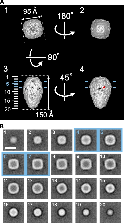
FIGURE 6.
Structural features of Orai1. A, surface representations of Orai1 viewed from four different Euler angles (α, β, γ): 1 (0, 180, -45), 2 (0, 0, -45), 3 (0, 90, -45), and 4 (0, 90, 0). The molecular mass enclosed by the isosurface is 210 kDa, corresponding to 149% of the tetrameric Orai1 protein. Protein is displayed in bright shades. Two blue lines, ∼30 Å apart in panels 3 and 4, indicate the putative position of the lipid bilayer. A red arrowhead in panel 4 indicates one of inverted-V-shaped orifices in the cytoplasmic domain. B, horizontal sections parallel to the membrane plane. Sections at 7.7-Å intervals through the molecule are indicated by numbers 1–20 on the left side of panel A3. The internal structure of the Orai1 molecule is sparse. Scale bar represents 100 Å.

