FIGURE 5.
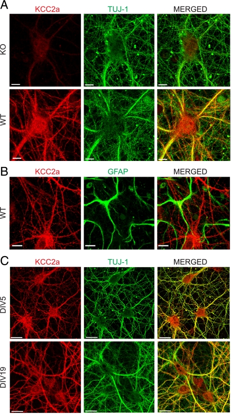
Localization of the KCC2a isoform in cultured neurons. Shown are maximum intensity projections of confocal optical images. A, hippocampal neurons, derived from E17 WT or KCC2 null mutant (KO) mice and cultured for 11 days in vitro (DIV11), were double-immunostained (see “Experimental Procedures”) with antibodies against KCC2a (red) and neuron-specific β-III tubulin (TUJ-1) (green). WT and KO cultures were treated in parallel, and the same settings were applied during the imaging procedures, thus allowing a direct comparison of the KCC2a staining in WT and KO neurons. KCC2a antibody revealed a strong KCC2a protein expression in WT neurons, whereas only residual background staining was found in KO neurons. B, DIV11 hippocampal cultures from WT mice were double-stained with antibodies against KCC2a (red) and GFAP (green). Non-overlapping patterns revealed by these antibodies confirmed an exclusively neuron-specific expression of the KCC2a protein. C, mouse hippocampal neuronal cultures, prepared similar to A and cultured for 5 or 19 days, were double-immunostained with KCC2a (red) and TUJ-1 (green) antibodies. KCC2a expression was exclusively neuronal at all time points analyzed. KCC2a immunostaining was present in cell bodies and dendritic shafts already at DIV5 and remained similar in DIV19 neurons. Scale bar is 10 μmin A and B and 20 μmin C.

