FIGURE 6.
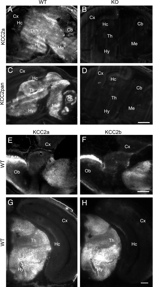
KCC2a and KCC2b protein isoforms have a similar pattern of expression in E18 mouse brain. Sagittal (A–D) or coronal (E–H) cryosections of E18 WT (A, C, and E–H) or KCC2 null mutant (KO, B and D) mice were analyzed by immunohistochemistry (see “Experimental Procedures”) with KCC2a (A, B, E, and G), KCC2pan (C and D), and KCC2b (F and H) antibodies. Immunoreactivity for KCC2a was widely distributed in non-cortical brain areas (A) of wild-type mice and was absent in the KCC2 null mutant littermates (B). The KCC2pan antibody produced a pattern similar to KCC2a antibody in wild-type mice (C) and no staining in sections from KCC2 null mutant embryos (D). Immunostaining of adjacent sections with the KCC2a and KCC2b antibodies revealed a similar distribution of the two KCC2 isoforms in the olfactory bulb (compare E and F) and thalamus (compare G and H). All three KCC2 antibodies detected only a weak signal in the cerebral cortex and hippocampus. The signal in the skin (asterisk in A and G) was unspecific. Cb, cerebellum; Cx, neocortex; Hc, hippocampus; Hy, hypothalamus; Me, medulla; Ob, olfactory bulb; Th, thalamus. Scale bar is 1 m min A–D; 0.5 mm in E and F, and 0.2 mm in G and H.

