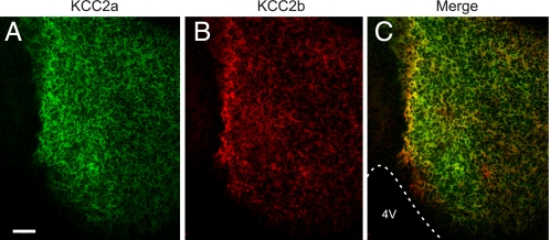
FIGURE 7.
KCC2a and KCC2b are colocalized in E18 mouse brain neurons. Shown are confocal optical section images of a coronal cryosection through E18 mouse midbrain double-stained with the KCC2a (green) and KCC2b (red) antibodies (see “Experimental Procedures”). C is the merged image of A and B. Most neurons in this area coexpress KCC2a and KCC2b, although the distribution is not identical. 4V, fourth ventricle. Scale bar is 50 μm.

