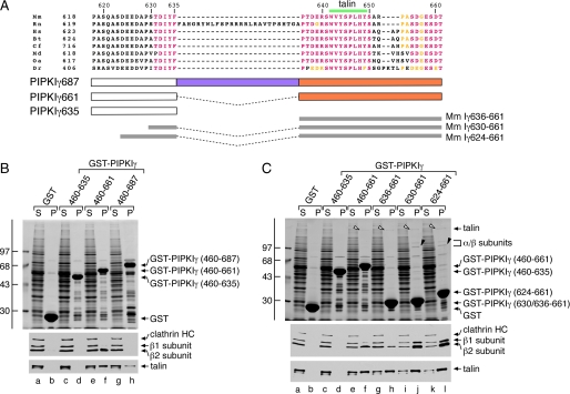
FIGURE 6.
Differing partner binding properties of the three PIPKIγ splice variants. A, primary sequence alignment of the C-terminal segment of PIPKIγ isoforms from various species: murine (Mm; NCBI accession number NP_032870.1), rat (Rn; NP_001009967.2), human (Hs; NP_036530.1), bovine (Bt; XP_585653.4), feline (Cf; XP_542172.2), opossum (Md; XP_001363745.1), platypus (Oa; XP_001511349.1), and zebrafish (Dr; XP_683392.3). Identical residues are colored pink, and conservatively substituted residues are yellow. The location of the talin-binding motif is indicated above, and the GST fusion proteins spanning the junction between the PIPKIγ635 and -661 isoforms tested is indicated below. B, ∼200 μg of GST (lanes a and b), GST-PIPKIγ-(460-635) (lanes c and d), GST-PIPKIγ-(460-661) (lanes e and f), or GST-PIPKIγ-(460-687) (lanes g and h) immobilized on glutathione-Sepharose was incubated with rat brain cytosol as indicated. After centrifugation, aliquots of 1.5% of each supernatant (S) and 10% of each washed pellet (P) were resolved by SDS-PAGE and either stained with Coomassie Blue or transferred to nitrocellulose. Portions of the blots were probed with anti-clathrin heavy chain (HC) mAb TD.1 and anti-β1/β2 subunit mAb 100/1 or with anti-talin mAb 8d4, and only the relevant portions are shown. C, ∼200 μg of GST (lanes a and b), GST-PIPKIγ-(460-635) (lanes c and d), GST-PIPKIγ-(460-661) (lanes e and f), GST-PIPKIγ-(636-661) (lanes g and h), GST-PIPKIγ-(630-661) (lanes i and j), or GST-PIPKIγ-(624-661) (lanes k and l) immobilized on glutathione-Sepharose was incubated with rat brain cytosol as indicated. After centrifugation, aliquots of 1.5% of each supernatant(S) and 10% of each washed pellet (P) were resolved by SDS-PAGE and either stained with Coomassie Blue or transferred to nitrocellulose. Portions of the blots were probed with anti-clathrin heavy chain (HC) mAb TD.1 and anti-β1/β2 subunit mAb 100/1 or anti-talin mAb 8d4, and only the relevant portions are shown. The position of talin (open arrowheads) and the AP-2 β2 and αC subunits (black arrowheads) on the stained gel is shown.

