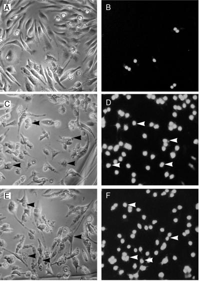
Figure 2.
Macrophages are killed after microinjection of SipB. Every macrophage in the field was microinjected with purified GST (A and B, same field), GST–IpaB (C and D, same field), or GST–SipB (E and F, same field) and then stained with PI in PBS without fixation. Apoptotic cells were then scored for PI uptake and cellular morphology. Many more cells are positive for PI uptake and apoptotic cellular alterations in GST–SipB (E and F) and GST–IpaB (C and D) injected cells than found in GST-injected cells (A and B). Arrowheads highlight some of the cells undergoing apoptosis. See text for quantification of the levels of apoptosis.

