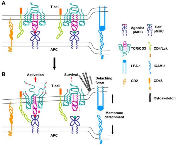
Figure 1.
The schematics of the receptor deformation model of TCR triggering. A) Interaction of pMHCs and TCRs at the close membrane-membrane contact site of the T cell/APC interface. Large adhesion molecules such as LFA-1 and ICAM-1 bring together the cell membranes, which are further aligned by small adhesion molecules such as CD2 and CD48, to a distance permitting pMHC-TCR interaction. The interaction between agonist pMHC and TCR per se does not initiate TCR signaling. B) Forces generated from the cytoskeleton, which drive active T cell movement relative to APC and T cell morphological changes, detach the T cell plasma membrane from the APC. Part of the force is delivered to the TCR/CD3 complex through pMHC-TCR binding. Binding between agonist pMHC and TCR is strong enough to deliver a force that deforms the TCR/CD3 complex to a conformation or configuration that can initiate an activation signal. Weak binding between self-pMHC and TCR breaks before such a force can be delivered. Such a binding could, however, deliver a weak force that causes minor receptor deformation, leading to a survival signal. The detaching force could also deform the MHC molecule to a conformation that binds coreceptors with higher affinity, or directly deform the coreceptors to alter the activity of associated tyrosine kinase Lck.

