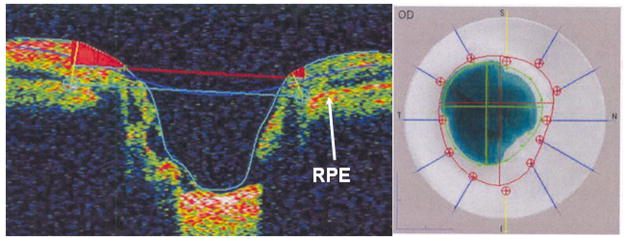
Figure 4.
Time domain optical coherence tomography (Stratus OCT3, Carl Zeiss Meditec Inc, Dublin, CA) imaging of the right optic nerve of a 52-year-old white man with physiologic cupping. Left image, This is the vertical cross-section through the optic nerve head. The reference plane (upper horizontal line) 150 μm above the retinal pigment epithelium (RPE) divides the neuroretinal rim above from the cup below. Right image, The cup is delimited by the inner circle and the disc border by the outer dotted circle.

