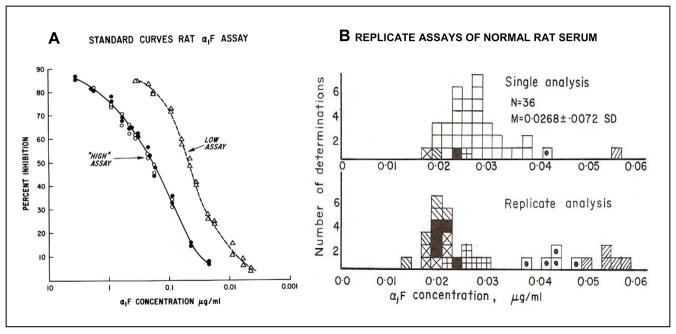
Figure 2. Radioimmunoassay (RIA) for rat AFP.
A. Standard curves for the “high” and the “low” RIA. The low assay was performed by pre-incubation of the unknown or the known standard with the anti-AFP before addition of the radio-labeled AFP. B. The top figure shows the range of AFP levels in normal rat serum. The bottom figure shows results of replicated assays for selected samples for normal rat serum. Each symbol represents the assays for a given serum.
Modified from Sell and Gord [20].

