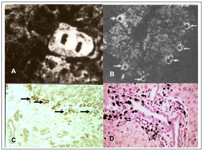
Figure 6. Cellular localization of AFP after various models of liver injury.
A. Immunofluorescence labeling of AFP in mitotic hepatocyte after partial hepatectomy [45]. B. Immunofluorescence labeling of AFP in pre-mitotic hepatocyte after CCl4 induced central injury [46]. C. Immunoperoxidase labeling of AFP in transitional oval cells after periportal injury induced by cocaine [59]. D. Tritiatiate thymidine labeling of oval duct cells and periductular cells after CCl4 injury and treatment with N-2-actylaminofluorene [55].

