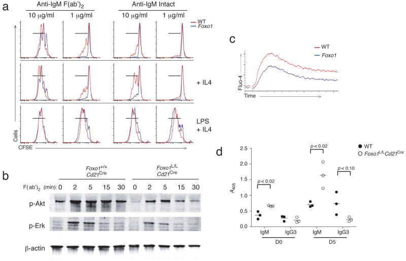
Figure 6. Foxo1L/LCd21Cre B cells have impaired responses to anti-IgM stimulation in vitro, but intact antibody responses to TI-2 antigens.
(a) Representative FACS plots of proliferation measurements of purified splenic B cells from Foxo1+/+Cd21Cre (red) and Foxo1L/LCd21Cre (blue) mice following CFSE labeling and stimulation (3 days) with 1 μg/mL or 10 μg/mL of either anti-IgM F(ab′)2 or intact anti-IgM in the presence or absence of IL-4; or stimulated with LPS and IL-4. CFSE partitioning was analyzed by FACS. (b) Immunoblot analysis of phospho-Akt and phospho-Erk from anti-IgM F(ab′)2 stimulated splenic B cells. (c) Calcium flux by purified splenic B cells from Foxo1+/+Cd21Cre (red) and Foxo1L/LCd21Cre (blue) mice loaded with Fluo-4 and stimulated with 10 μg/mL anti-IgM F(ab′)2. (d) ELISA to detect TNP-specific IgM and IgG3 present in the serum of Foxo1+/+Cd21Cre (open circles) and Foxo1L/LCd21Cre (filled circles) mice before and 5 days after IP immunization with 10 μg TNP-Ficoll in PBS. FACS profile and in vitro stimulation is representative of 3 independent experiments. ELISA data is representative of 2 independent experiments with 3 mice per group.

