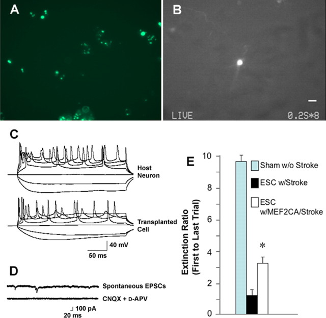
Figure 7.
MEF2CA-ESC-derived neuronal progenitor cells injected into ischemic mouse brain manifest electrophysiological properties of integrated neurons and improve behavior. A, MEF2CA-expressing cells labeled with Qtracker 525 (Invitrogen) before injection. B, Representative transplanted cell containing quantum dots visualized live using a FITC filter in an acute cortical brain slice obtained 6 weeks after injection into the striatum of the mouse stroke model. Careful inspection reveals the presence of long fluorescent processes emanating from the cell body. Scale bar, 10 μm. Similar cells were recorded from in the electrophysiological traces shown below. For this purpose, we selected engrafted cells that were isolated from others to carefully characterize their potential integration into the surrounding endogenous neuronal network by assessing synaptic activity. C, During whole-cell recording in the current-clamp mode, both endogenous unlabeled neurons (top trace) and transplanted green cells (bottom trace) displayed similar trains of action potentials, which were elicited by injection of depolarizing current, and passive voltage responses to hyperpolarizing current (n = 6 cells). D, Under voltage clamp, an engrafted (green) cell in a cortical slice was held at −60 mV, and sEPSCs were observed (top trace). These synaptic currents were completely blocked by perfusion in a combination of 10 μm CNQX and 50 μm d-APV (bottom trace; n = 3 cells). E, Three months after tMCAOR and injection of either MEF2CA/EGFP-ESC-derived NPCs or EGFP-expressing progenitor cells, or sham surgery, mice underwent a standard neurobehavioral battery of fear conditioning and were then tested for extinction of the fear response (n = 10 for each group; *p < 0.01). Error bars indicate SEM.

