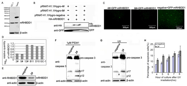
Figure 2.
Apoptosis in mRHBDD1 knock-down GC-1 cells. A. mRHBDD1 protein levels in GC-1 cells and mouse testis were examined by Western blotting; the same blot was immunoblotted with anti-β-actin antibody as internal control. Protein (30 μg) was separated on a 12% polyacrylamide gel as described in the Methods section. B and C. Screening for effective RNAi plasmids against mRHBDD1. Western blotting indicated that pRNAT-H1.1/Hygro-4# and 6# were both candidates; pRNAT-H1.1/Hygro-6# was selected. GFP was a loading control. D and E. Identification of stable mRHBDD1 knockdown GC-1 cell clones by Real-Time PCR and Western blotting. Clone 2# was selected for the following experiments. F and G. Western blot analysis of caspase 3 cleavage in 2# and negative control GC-1 cells after PS341 or UV treatment. Control cells were subcultured from a selected stable GC-1 cell clone expressing a control negative shRNA. H. FACS assay to analyse apoptosis in 2# and negative control GC-1 cells after UV irradiation. The percentage of apoptotic cells (% of total cells) was determined using the program EXPO32-ADC and shown as a bar chart. * (p < 0.05)

