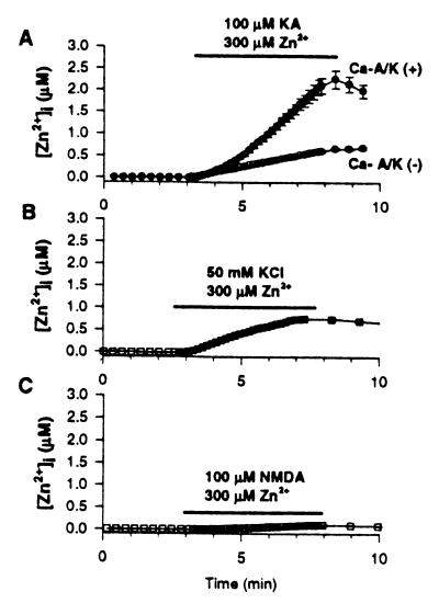
Figure 2.
Kainate + Zn2+ exposures trigger high Δ[Zn2+]i in Ca-A/K(+) neurons. Cortical cultures were loaded with Newport green and, after recording baseline fluorescence, were exposed for 5 min to 300 μM Zn2+ with 100 μM kainate (+10 μM MK-801; A); with 50 mM K+ (+10 μM MK-801, 10 μM NBQX; B); or with 100 μM NMDA (+10 μM NBQX; C). Ca-A/K(+) neurons are shown only in A, as control experiments showed no difference in Δ[Zn2+]i between Ca-A/K(+) and Ca-A/K(−) neurons after high-K+ or NMDA exposures. Traces show mean (±SEM) of 13 Ca-A/K(+) neurons (A) and ≥44 of either Ca-A/K(−) (A) or total neurons (B, C) from one experiment representative of ≥5. Note the particularly high kainate-triggered Δ[Zn2+]i in Ca-A/K(+) neurons and lower Δ[Zn2+]i in all neurons after high-K+ or NMDA exposures.

