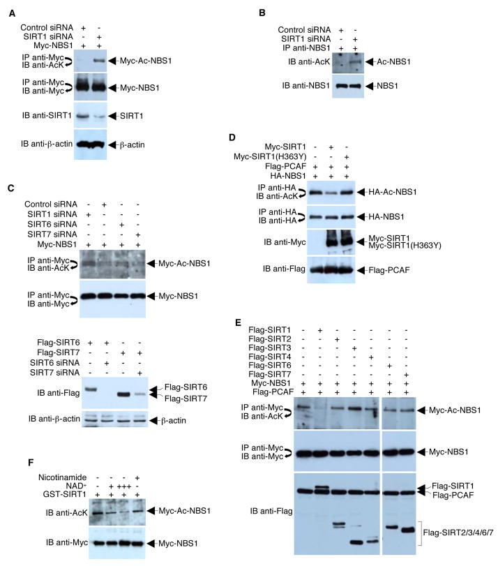
Figure 3. SIRT1 Deacetylates NBS1.
(A) 293T cells were co-transfected with equal amounts (4 μg) of Myc-NBS1 plasmid and either a plasmid encoding pBS/U6-SIRT1 or control siRNA. Western blots were performed with the indicated antibodies to assess the levels of SIRT1, β-actin, total Myc-NBS1, and acetylated Myc-NBS1. (B) 293T cells were infected with either adenovirus that expresses control siRNA or adenovirus that expresses SIRT1 siRNA. Western blots were performed with the indicated antibodies to assess the acetylation of endogenous NBS1 and NBS1 immunoprecipitation efficiency. (C) Upper panels, 293T cells were co-transfected with equal amounts (4 μg) of Myc-NBS1 plasmid and either a plasmid encoding SIRT1 siRNA, SIRT6 siRNA, SIRT7 siRNA, or control siRNA. Western blots were performed with the indicated antibodies to assess the levels of total Myc-NBS1 and acetylated Myc-NBS1. Bottom panels, similar experiments were performed with over-expression of Flag-SIRT6 and Flag-SIRT7 to show that SIRT6 and SIRT7 siRNAs are functional. D) 293T cells were co-transfected with plasmids that express HA-NBS1, Flag-PCAF, and either a wild-type or a catalytically-defective Myc-SIRT1. Acetylation of HA-NBS1 and all protein levels were determined with direct Western blotting or immunoprecipitations followed by Western blotting using the indicated antibodies. (E) 293T cells were co-transfected with plasmids that express Myc-NBS1, Flag-PCAF, and different Flag-tagged SIRTs. Acetylation of Myc-NBS1 and all protein levels were determined with direct Western blotting or immunoprecipitations followed by Western blotting using the indicated antibodies. (F) 293T cells were transfected with Myc-NBS1 and Flag-PCAF. Anti-Myc immunoprecipitates were incubated with recombinant GST-SIRT1 in the presence or absence of NAD+ (+, 1mM; +++, 10 mM) and nicotinamide (10 mM) at 30°C for 1 h. Western blot analysis of acetylated Myc-NBS1 was then performed using anti-acetyl-lysine (AcK) antibodies.

