
Figure 4.
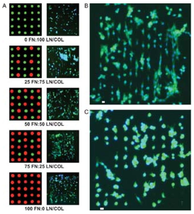
C2C12 myoblast adhesion and spreading on two component protein arrays. A) Fluorescence images of cell adhesion and spreading on the composition gradient of fibronectin (FN) and lamnin/collagen (LN/COL). B) Fluorescence image of aligned cell adhesion on the protein array with a composition of 25/75 (FN/(LN/COL) and a spacing of 18 μm. C) Fluorescence image of cell adhesion on protein array with a composition of 25/75 (FN/(LN/COL) and a spot spacing of 24 μm. The scale bars in the Figure 4b and 4c represent 20 μm.