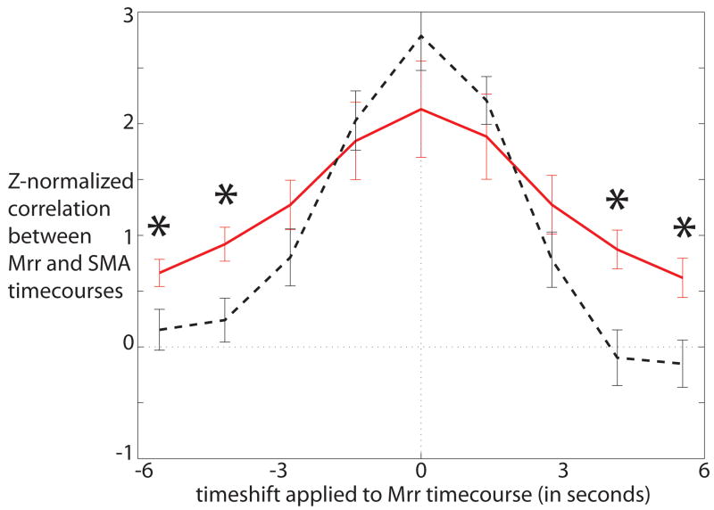
Figure 1.
Temporal cross-correlation between the functionally defined motor cortical reference region (Mrr) and the SMA during tics (red solid line) and during intentional movements in control subjects (black dashed line). Asterisks indicate differences between patients and controls at specific time-points as identified in the post-hoc t-tests. SMA is more active both prior to and after the motor cortical reference region during tics than it is during intentional movements.

