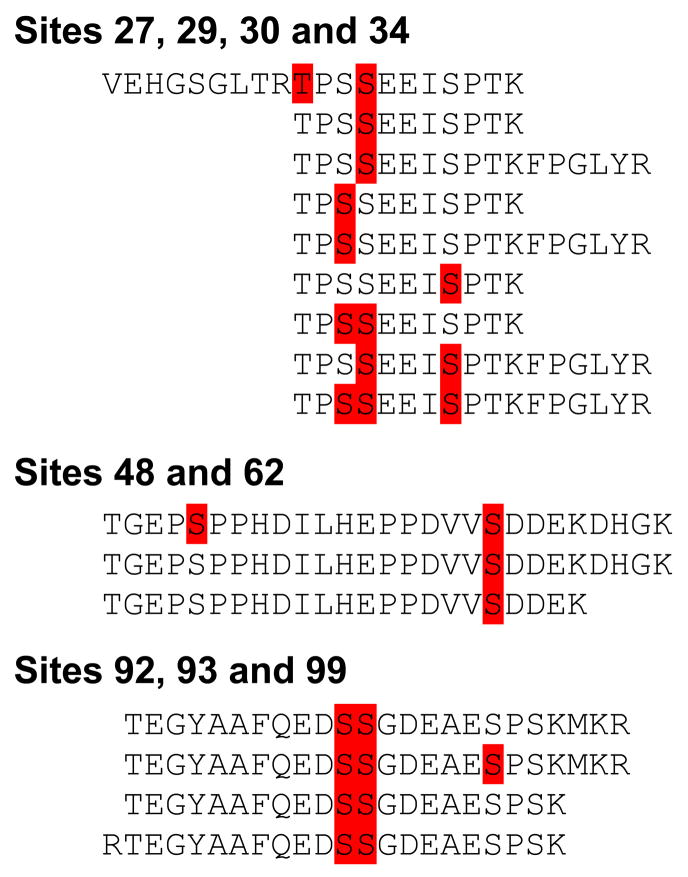
Figure 3.
In three different regions several peptides were phosphorylated at multiple sites. Detected phosphorylations are indicated in red; peptides detected are written out. In the first region, only one peptide was detected with phosphorylation at T27. Most S29, S30 and S34 phosphorylation patterns were detectable, with one exception. S29 was never phosphorylated together with S34 alone. In the second region, pS48 was only detected in peptides with pS62 peptides. No peptides were detected without S62 phosphorylation. The third region where more than one residue was detected to be phosphorylated contained the S92, S93, S99 sites. No peptides without S92 and S93 phosphorylation were detected. These observations are identical between the UMUC-3 and 293T cell lines.

