
Figure 1.
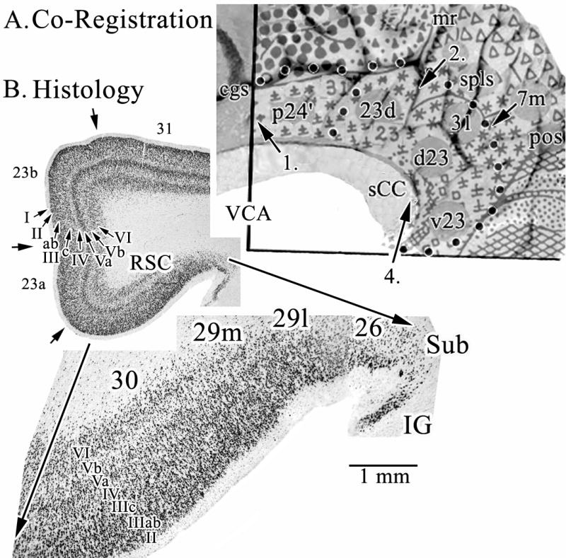
A. Co-registration of Brodmann’s map of posterior cingulate and precuneal cortices and a postmortem case for joint histological assessment. Stroked dots outline the border of cingulate cortex and four numbered arrows refer to locations of particular Brodmann areas discussed in the text. Brodmann’s actual numbers 23 and 31 are shown in the co-registration, while all other numbers refer to Vogt et al. (2005). The arrow at 4. points to the level of the coronal histological section taken for B. A critical issue at 4. is the manner in which Brodmann extended retrosplenial areas 29 and 30 onto the gyral surface where area 23 is located. RSC comprises the ventral bank of the cingulate gyrus in the callosal sulcus and is not exposed as suggested in his map. Abbreviations: cgs, cingulate sulcus; IG, indusium griseum; mr, marginal ramus of the cgs; pos, parieto-occipital sulcus; sCC, splenium of the corpus callosum; Sub, dorsal subiculum; VCA, vertical plane at the anterior commissure;