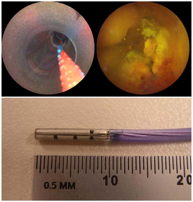
Figure 1.
SFE images during in vivo imaging in a pig bile duct using ERCP procedure, top-left shows the 4.2-mm working channel and guidewire and top-right shows a SFE image frame. Photograph of SFE probe with 9-mm rigid tip length and 1.2-mm overall diameter is the lower image. Reprinted from [7] Seibel, E.J., Melville, C.D., Johnston, R.S., et al., Bile duct imaging with ultrathin laser scanning catheterscope in a swine model, Gastrointest Endosc 2008;67(5):AB133–134, with permission from Elsevier.

