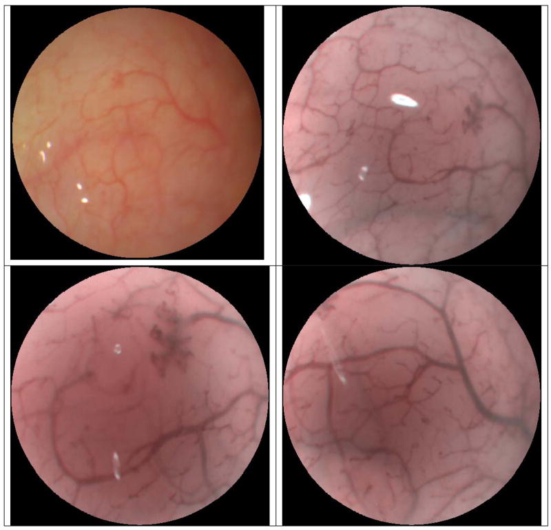
Figure 3.
TCE images showing video frame in normal operation (top-left) and subsequent video frames captured with Enhanced Spectral Imaging (ESI) feature applied (top-right). TCE w/ESI is demonstrated with increased magnification within the previous field of view (bottom-left), and in a new region of the human cheek (bottom-right).

