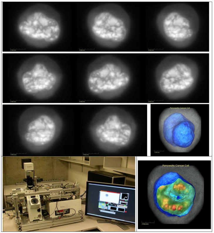
Figure 7.
Cell-CT instrument and transmission images of human pancreatic cancer cell (PaTu1, alcohol fixed, hematoxylin stained) shown in grayscale in series of different rotational perspectives. Same cell is visualized using Volview software with thresholding set to opacify the irregular nuclear envelope (blue) and highlight chromatin-dense bodies within slice of nucleus (red).

