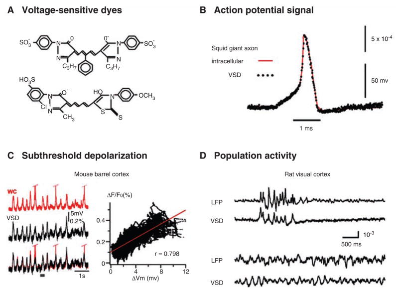
Fig. 1.
(A) Structural formula of commonly used voltage-sensitive dyes (VSD). (Top) RH482 (also known as NK3630), an absorption dye used for imaging neuronal activity in brain slices. (Bottom) RH1691, a fluorescent dye commonly used for imaging cortex in vivo. It is one of newly developed “blue” dyes, and it has small pulsation artifacts for imaging cortex in vivo (Shoham and others 1999). (Structural formulae for more dyes can be found at: http://square.umin.ac.jp/optical/optical/dye.html.) (B) Simultaneous VSD (dots) and intracellular (line) recordings from a squid giant axon. The axon is stained with an absorption dye. The two signals follow each other precisely, providing the first evidence that dye signal is membrane potential dependent. Note that although the linearity and temporal response of the dye signal are excellent, the amplitude of the dye signal is small, only about 0.1% change from the resting light level per 100 mV changes in membrane potential. (Reprint from Ross and others 1977, with permission from Springer.) (C, left) Simultaneous recordings of intra-cellular potential (whole-cell patch, red) from one cortical neuron and VSD signal (black) from the surrounding population of neurons. The cortex is stained with RH1691. Under anesthesia, cortical neurons undergo synchronized up and down states. The subthreshold membrane potential fluctuations are closely correlated with the VSD signal, but the spikes on individual neurons were not correlated with large deflections of the VSD signal. (C, right) The VSD signal is plotted against membrane potential changes, demonstrating a linear relationship. (Modified from Ferezou and others 2006, with permission from Elsevier.) (D) Simultaneous local field potential (LFP) and VSD recordings from the rat visual cortex, demonstrating the sensitivity of VSD imaging compared to that of LFP (microelectroencephalography). The top two traces show synchronized bursting activity recorded under 1.5% isoflurane anesthesia. The correlation is high between LFP and VSD signals. The bottom two traces were recorded later under lower anesthesia level. Both LFP and VSD signals show sleep-like oscillations but the correlation remains low. The poor correlation is unlikely attributed to the baseline noise, because the baseline noise of the analogous recording under higher anesthesia is low. (Reprint from Lippert and others 2007, with permission from the American Physiological Society.)

