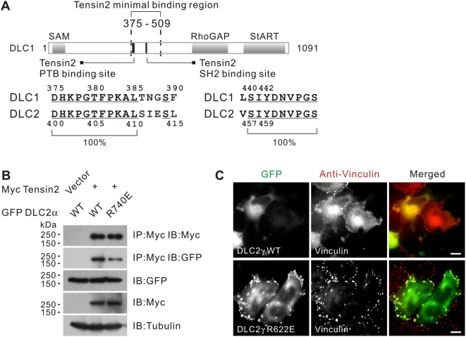
Figure 3. Conservation of the tensin2 PTB binding site in DLC1/2.
(A) Schematic showing the tensin2 SH2 and PTB binding sites in DLC1. Within the minimal tensin2 binding region 375–509, separate elements are involved in mediating tensin2 binding via PTB or SH2 mechanisms. These elements are well conserved in the DLC1 paralog, DLC2. Conserved residues were underlined. (B) Interaction between DLC2 and tensin2 in a RhoGAP-independent manner. HEK293T cells were transfected with Myc-tagged tensin2 and GFP-tagged DLC2 constructs as indicated. Cleared cell lysates were incubated with anti-Myc antibody to immunoprecipitate tensin2. DLC2 in the precipitates was detected by immunoblotting with anti-GFP antibody. (C) GFP-DLC2γ showed focal adhesion localization in a RhoGA-independent manner. Expression of GFP-DLC2γ induced severe cell shrinkage when expressed in SMMC-7721 cells. Focal adhesion localization of the DLC2γ RhoGAP mutant R622E was detected. The endogenous vinculin was visualized by anti-vinculin antibody. Scale bar = 10 µm.

