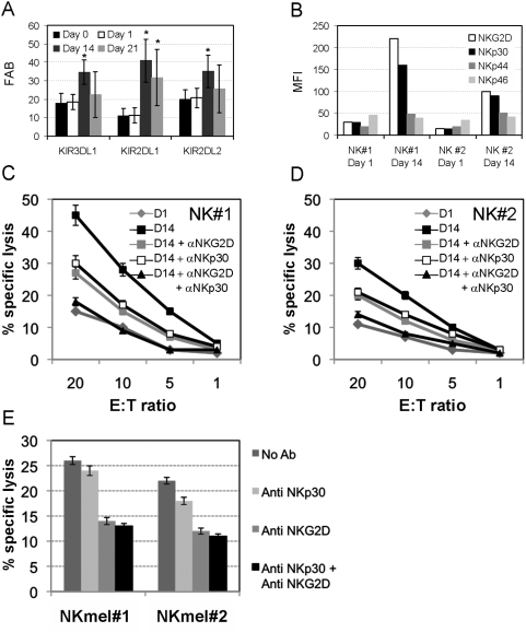
Figure 9. Enhanced expression of NKLRs correlates with improved cytotoxicity against melanoma cells.
(A) KIR expression during ex vivo expansion. All NK cultures were independently stained for the indicated KIRs. Expression of KIRs in each donor was normalized according to the corresponding expression on gated NK cells by the un-manipulated peripheral blood mononuclear cells (PBMC). Figure shows the average of normalized results on all healthy donors tested (N = 7). Error bars represent standard error. * denotes a statistical significance of P value<0.05. (B) The expression of each NKLR was tested on two representative bulk NK cultures on day 1 and day 14 of ex vivo expansion process, as indicated in the figure. Expression is presented as median fluorescence intensity (MFI). (C) NK#1 and (D) NK#2 cultures were tested in killing assays against Mel008 cells in various effector-to-target (E∶T) ratios. NKG2D or/and NKp30 on NK cultures #1 and #2 at day 14 were blocked by pre-incubation with mAb at a concentration of 2 µg/ml or 4 µg/ml, respectively. Figure shows a representative experiment. (E) NK cells with low expression of NKp30 were derived from melanoma patients (NKmel#1 and NKmel#2) and tested in killing assays against Mel008 cells in an E∶T of 20∶1. NKG2D or/and NKp30 were blocked as described above. Figure shows a representative experiment.

