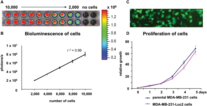
Figure 1. In vitro evaluation of MDA-MB-231-Luc2 cells.
(A) Cells were plated in a 96-well plate in quadruplicate, ranging from 10,000 to 2,000 cell/well. Wells with media only (no cells) were included as control. D-luciferin substrate was added to each well and the plate was imaged to estimate the bioluminescence of MDA-MB-231-Luc2 cells. (B) Correlation between bioluminescence and cell number per well was plotted as mean photons/s/well±SEM. (C) Fluorescence microscopy, showing that MDA-MB-231-Luc2 cells expressed EGFP. (D) 1×10³ MDA-MB-231 parental and MDA-MB-231-Luc2 cells were seeded into quadruplicate wells of 96-well plates and their proliferation rates determined by MTT assay. Data was plotted as mean relative growth±SEM.

