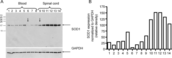
Figure 4 Immunoblotting analysis of SOD1 expression
(A) Western blot with arrow points to the samples from the proband. Protein lysates of white blood cells from family members heterozygous (lanes 1–4 and 7–8), wild-type (lane 5), and homozygous (lane 6) for the ΔG27/P28 deletion, and lumbar spinal cord extracts from the ΔG27/P28 homozygote (lane 9), familial amyotrophic lateral sclerosis (ALS) A4V case (lane 10), two sporadic ALS cases (lanes 11–12), Alzheimer disease case (lane 13), and corticobasal syndrome case (lane 14) immunoblotted against anti-SOD1 antibody. The membrane was reprobed with anti-GAPDH antibody to show equal loading. (B) Quantification of SOD1 protein expression based on immunoblotting band intensity (relative density normalized to GAPDH).

