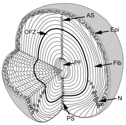
Fig. 1.
Cellular organization of the mouse lens. The lens consists of two cell types: epithelial cells (Epi) located at the anterior surface, and fiber cells (Fib), which comprise the remainder and majority of the tissue. At the lens equator, epithelial cells differentiate into fiber cells. As they differentiate, fibers become highly elongated. The tips of the elongating fibers converge at the anterior and posterior lens sutures (AS and PS, respectively). In cross section, the fiber cells have a flattened hexagonal profile with two broad faces (oriented parallel to the lens surface) and four narrow faces. Initially, all fiber cells are nucleated but, during differentiation, nuclei (N) and other organelles are degraded. As a result, the central region of the lens constitutes an organelle-free zone (OFZ). The innermost cells, termed primary fiber cells (PF), are formed during embryonic development and are less regular in shape and arrangement than the other fiber cells.

