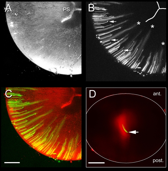
Fig. 2.
The large molecule diffusion pathway (LMDP) is established in elongating fiber cells and persists in mature fiber cells. A-C. Maximum intensity projections of the posterior lens hemisphere from a 33-day-old Cre-ER™;Z/EG mouse, 10 days after tamoxifen treatment. The lens was incubated in FM4-64 to visualize the membrane architecture. (A) FM4-64 staining reveals the regular arrangement of the fiber cells and the position of the Y-shaped posterior suture (PS). (B) In short, newly differentiated cells near the periphery, GFP is restricted to the cytoplasm of expressing cells (arrows). As fiber elongation proceeds, GFP diffuses from expressing cells into the cytoplasm of neighboring cells (*). Note that intercellular diffusion commences before cells reach the suture (i.e. in actively elongating fiber cells) (see Fig. 1). (C) Co-visualization of GFP (green) and FM4-64 fluorescence (red). (D) Wild-type mouse lens 10 minutes (green) or 2 days (red) after injection of Alexa Fluor 488-dextran (10 kDa) into a fiber cell located in the center of the lens (arrow). Note the extensive intercellular spread of tracer over the 2 day incubation period. Scale bars: 250 μm.

